
Welcome to the ultimate guide to understanding the heart’s anatomy and function through the lens of heart diagram worksheet PDF answers. This comprehensive resource will provide you with a detailed analysis of the heart diagram worksheet, highlighting key features and their significance.
Delve into the role of the heart in the circulatory system and explore common heart conditions and disorders. Prepare to embark on an educational journey that will leave you with a profound understanding of this vital organ.
The human heart is a remarkable organ that plays a central role in maintaining life. Its intricate structure and function are essential for pumping blood throughout the body, delivering oxygen and nutrients to tissues and organs. To gain a deeper understanding of the heart’s anatomy and function, it is important to examine heart diagrams and worksheets, which provide visual representations of this complex organ.
Heart Anatomy Overview
The heart is a vital organ responsible for pumping blood throughout the body, supplying oxygen and nutrients to tissues and removing waste products. It is located in the center of the chest cavity, slightly to the left of the midline, and is protected by the rib cage.
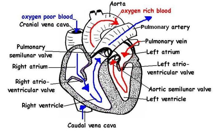
The heart has a muscular structure and is divided into four chambers: two atria (upper chambers) and two ventricles (lower chambers). The atria receive blood from the body and the ventricles pump blood out to the body. Between the atria and ventricles are valves that prevent backflow of blood.
The major blood vessels connected to the heart are the aorta, which carries oxygenated blood away from the heart to the body, and the vena cava, which returns deoxygenated blood to the heart from the body.
Chambers of the Heart
The heart has four chambers: two atria (upper chambers) and two ventricles (lower chambers). The right atrium receives deoxygenated blood from the body through the vena cava. The right ventricle pumps this blood to the lungs through the pulmonary artery, where it is oxygenated.
The left atrium receives oxygenated blood from the lungs through the pulmonary vein. The left ventricle pumps this blood to the body through the aorta.
Valves of the Heart, Heart diagram worksheet pdf answers
The heart has four valves that prevent backflow of blood. The tricuspid valve is located between the right atrium and right ventricle. The pulmonary valve is located between the right ventricle and pulmonary artery. The mitral valve (also known as the bicuspid valve) is located between the left atrium and left ventricle.
The aortic valve is located between the left ventricle and aorta.
Major Blood Vessels
The major blood vessels connected to the heart are the aorta, which carries oxygenated blood away from the heart to the body, and the vena cava, which returns deoxygenated blood to the heart from the body. The aorta is the largest artery in the body and branches into smaller arteries that supply blood to all tissues.
The vena cava is formed by the union of two large veins, the superior vena cava and the inferior vena cava, which collect deoxygenated blood from the upper and lower body, respectively.
Heart Diagram Worksheet Analysis
The heart diagram worksheet PDF provides a visual representation of the heart’s anatomy, enabling students to identify and understand the various components of this vital organ. The diagram includes detailed illustrations of the heart’s chambers, valves, blood vessels, and surrounding structures.
By analyzing the heart diagram, students can gain a deeper understanding of the heart’s structure and function. The worksheet includes key features such as the four chambers (right atrium, right ventricle, left atrium, and left ventricle), the atrioventricular valves (tricuspid and mitral valves), and the semilunar valves (aortic and pulmonary valves).
Chambers of the Heart
The four chambers of the heart are responsible for receiving, pumping, and distributing blood throughout the body. The right atrium receives deoxygenated blood from the body, which then flows into the right ventricle. The right ventricle pumps the deoxygenated blood to the lungs, where it becomes oxygenated.
The oxygenated blood returns to the heart via the left atrium and is then pumped into the left ventricle. The left ventricle pumps the oxygenated blood to the rest of the body via the aorta.
Valves of the Heart, Heart diagram worksheet pdf answers
The heart valves play a crucial role in ensuring the proper flow of blood through the heart. The atrioventricular valves (tricuspid and mitral valves) prevent blood from flowing back into the atria when the ventricles contract. The semilunar valves (aortic and pulmonary valves) prevent blood from flowing back into the ventricles when the heart relaxes.
Blood Vessels of the Heart
The heart diagram also includes illustrations of the major blood vessels associated with the heart. The aorta is the main artery that carries oxygenated blood away from the heart to the rest of the body. The pulmonary artery carries deoxygenated blood from the heart to the lungs.
The pulmonary veins carry oxygenated blood from the lungs back to the heart. The coronary arteries supply blood to the heart muscle itself.
Surrounding Structures of the Heart
The heart diagram may also include illustrations of the surrounding structures that support the heart’s function. These structures include the pericardium, which is a sac that surrounds the heart, and the diaphragm, which is a muscle that separates the chest cavity from the abdominal cavity.
Heart Functions and Blood Flow
The heart is the central organ of the circulatory system, responsible for pumping oxygenated blood throughout the body and removing deoxygenated blood.
The heart is divided into four chambers: two atria (upper chambers) and two ventricles (lower chambers). The right atrium receives deoxygenated blood from the body, which then flows into the right ventricle. The right ventricle pumps the deoxygenated blood to the lungs, where it is oxygenated.
The oxygenated blood returns to the heart via the left atrium and flows into the left ventricle. The left ventricle pumps the oxygenated blood to the body.
Role of Heart Valves
The heart valves prevent the backflow of blood within the heart. The tricuspid valve is located between the right atrium and right ventricle, the pulmonary valve is located between the right ventricle and pulmonary artery, the mitral valve (also known as the bicuspid valve) is located between the left atrium and left ventricle, and the aortic valve is located between the left ventricle and aorta.
Heart Conditions and Disorders: Heart Diagram Worksheet Pdf Answers
Heart conditions and disorders encompass a wide range of diseases and abnormalities that affect the heart’s structure, function, and blood flow. These conditions can vary in severity, from mild and manageable to life-threatening. Understanding the symptoms, causes, and treatments of common heart conditions is crucial for maintaining cardiovascular health.
Heart conditions can be classified into several categories, including:
Coronary Artery Disease
- Coronary artery disease (CAD) is a condition in which the arteries that supply blood to the heart become narrowed or blocked by plaque buildup. This can lead to chest pain (angina), heart attack, or sudden cardiac death.
- Symptoms of CAD can include chest pain, shortness of breath, fatigue, and palpitations.
- Treatment for CAD typically involves lifestyle changes, medications, and in some cases, surgery.
Heart Failure
- Heart failure occurs when the heart is unable to pump enough blood to meet the body’s needs. This can be caused by various factors, including CAD, high blood pressure, or diabetes.
- Symptoms of heart failure can include shortness of breath, fatigue, swelling in the legs and feet, and rapid or irregular heartbeat.
- Treatment for heart failure aims to improve heart function and reduce symptoms, often involving medications, lifestyle changes, and in severe cases, heart transplant.
Arrhythmias
- Arrhythmias are disorders of the heart’s electrical system, which can cause the heart to beat too fast, too slowly, or irregularly.
- Symptoms of arrhythmias can include palpitations, chest pain, dizziness, or fainting.
- Treatment for arrhythmias may involve medications, implantable devices (such as pacemakers or defibrillators), or ablation procedures.
Valvular Heart Disease
- Valvular heart disease refers to conditions affecting the heart valves, which control the flow of blood through the heart. These conditions can include valve stenosis (narrowing) or regurgitation (leaking).
- Symptoms of valvular heart disease can vary depending on the affected valve and the severity of the condition, but may include shortness of breath, fatigue, chest pain, or palpitations.
- Treatment for valvular heart disease may involve medications, lifestyle changes, or surgical repair or replacement of the affected valve.
Congenital Heart Defects
- Congenital heart defects are structural abnormalities of the heart that are present at birth. These defects can range from mild to severe and may require medical intervention.
- Symptoms of congenital heart defects can vary depending on the type and severity of the defect, but may include shortness of breath, cyanosis (bluish skin), feeding difficulties, or developmental delays.
- Treatment for congenital heart defects may involve medications, surgeries, or other interventions, depending on the specific defect.
Educational Resources
The heart is a complex organ that plays a vital role in the human body. There are numerous educational resources available online and offline to help people learn more about the heart and its functions.
These resources can be beneficial for students, teachers, healthcare professionals, and anyone interested in gaining a better understanding of the heart.
Websites
- American Heart Association: https://www.heart.org
- National Heart, Lung, and Blood Institute: https://www.nhlbi.nih.gov
- Mayo Clinic: https://www.mayoclinic.org/diseases-conditions/heart-disease/symptoms-causes/syc-20353142
Articles
- “The Heart: A Vital Organ”by the American Heart Association: https://www.heart.org/en/health-topics/heart-attack/understanding-your-risk/the-heart-a-vital-organ
- “How the Heart Works”by the National Heart, Lung, and Blood Institute: https://www.nhlbi.nih.gov/health-topics/how-heart-works
- “Heart Disease: Symptoms, Causes, and Treatment”by the Mayo Clinic: https://www.mayoclinic.org/diseases-conditions/heart-disease/symptoms-causes/syc-20353142
Videos
- “The Heart and How It Works”by Khan Academy: https://www.khanacademy.org/science/ap-biology/human-biology/circulatory-system/a/the-heart
- “How the Heart Pumps Blood”by the National Heart, Lung, and Blood Institute: https://www.nhlbi.nih.gov/health-topics/how-heart-pumps-blood
- “Heart Disease: What You Need to Know”by the American Heart Association: https://www.heart.org/en/health-topics/heart-disease/understanding-your-risk/heart-disease-what-you-need-to-know
Commonly Asked Questions
What is the function of the heart?
The heart’s primary function is to pump blood throughout the body, delivering oxygen and nutrients to tissues and organs.
What are the four chambers of the heart?
The heart has four chambers: the right atrium, right ventricle, left atrium, and left ventricle.
What are the major blood vessels connected to the heart?
The major blood vessels connected to the heart include the aorta, pulmonary artery, superior vena cava, and inferior vena cava.
